Revision Rhinoplasty
Dr. Naficy specializes in corrective surgery (revision rhinoplasty) for situations where the outcome from an initial rhinoplasty surgery was less than ideal. There are a number of reasons why patients seek a secondary rhinoplasty procedure. In certain situations, the initial procedure may not have fully corrected the undesirable features of the nose (for example there is still a partial hump or the nose is still too large). In other situations, the initial rhinoplasty may have actually resulted in certain undesirable features (such as polybeak deformity, alar retraction, saddle deformity).
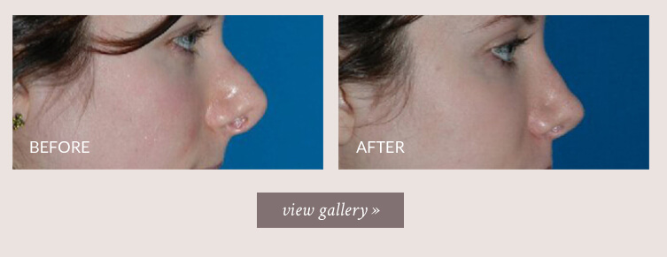
For example, the young woman below had a pollybeak deformity (polly beak) or supratip deformity of her nose resulting from her original rhinoplasty procedure years ago. Her nose had a beak-like hump on the profile and her tip was round and full. She underwent revision rhinoplasty with septal cartilage graft to help correct her problems and to obtain a more natural and aesthetically pleasing contour to her nose. In revision rhinoplasty, often a very small change, in the proper location, can make a big difference in the overall look and feel of the nose.
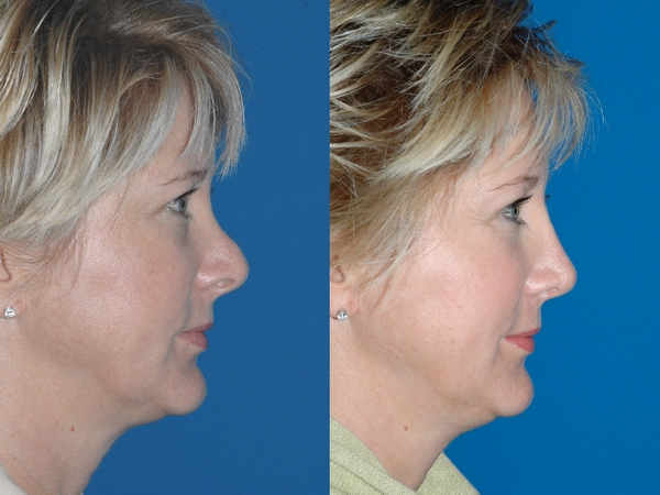
Revision rhinoplasty using septal cartilage graft by Dr. Sam Naficy. * Individual results may vary.
Dr. Naficy's practice has been 100% devoted to rhinoplasty and facial plastic surgery for the past 18 years. This is why Seattle doctors and patients have voted Dr. Naficy one of the top plastic surgeons for the face. Patients from the entire globe seek Dr. Naficy's expertise and he has many patients flying in for revision rhinoplasty surgery both nationally and internationally.
Revision Rhinoplasty Photo Gallery
Here you will find a sampling of representative before and after images of revision rhinoplasty and complex secondary rhinoplasty procedures performed by Dr. Sam Naficy. Click on any of the thumbnails to enter the slide show. All revision rhinoplasty and other facial plastic surgery were performed by Dr. Sam Naficy. The text accompanying the photos describes the details of the procedures performed.
* Individual results may vary.
Revision Rhinoplasty for Alar Retraction
Alar retraction is one of the common reasons for revision rhinoplasty. Alar retraction is present when the nostrils are pulled up due to inadeqaute support and there is excessive visibility of the inside of the nose. Alar retraction typically results from removal of excessive amounts of cartilage or failure to add support to the nostrils during rhinoplasty. Correction of alar retraction almost always requires cartilage grafting to provide support to the nostrils margins. This cartilage can come from the nose itself (septal cartilage), from the ear (auricular cartilage), or from the rib (costal cartilage).
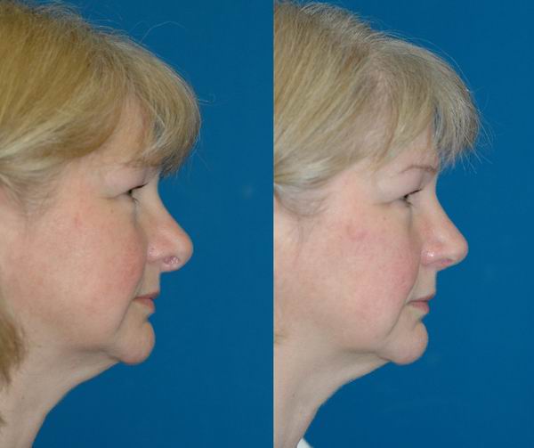
Revision rhinoplasty for alar retraction using septal cartilage and rib cartilage graft by Dr. Sam Naficy. * Individual results may vary.
Revision Rhinoplasty for Polybeak Deformity
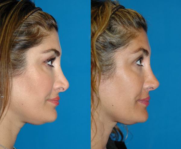
The pollybeak deformity is also known as the polly beak deformity, parrot beak deformity, or supratip deformity and is one of the common revision rhinoplasty problems. As the name implies, the pollybeak deformity gives a 'parrot-beak' shape to the profile of the nose. In profile, there is a hump-like fullness and heaviness just above the tip and the upper part of the ridge appears scooped. Pollybeak noses can at times have functional (breathing) problems. The pollybeak nose is typically the result of previous rhinoplasty surgery where excessive amounts of the bridge of the nose were removed. When the bridge is overly reduced, especially in someone with thicker skin, the excess skin tends to sag down and gather over the tip (in the supratip area) creating this deformity. The tip of the nose is also often not adequately supported which contributes to the problem. A pollybeak deformity is treated by adding (yes adding, not removing!) cartilage to rebuild and support the bridge and tip of the nose. The cartilage that is used for grafting typically comes from the septum or the rib. Choice of the grafting material depends on the severity of the problem with rib cartilage being able to provide more dramatic changes due to its strength and availability whereas septal cartilage is often available in smaller quantities and are less structurally strong. The skin will stretch to accommodate the newly framework of the nose and the fullness and heaviness in the supratip area will be improved.
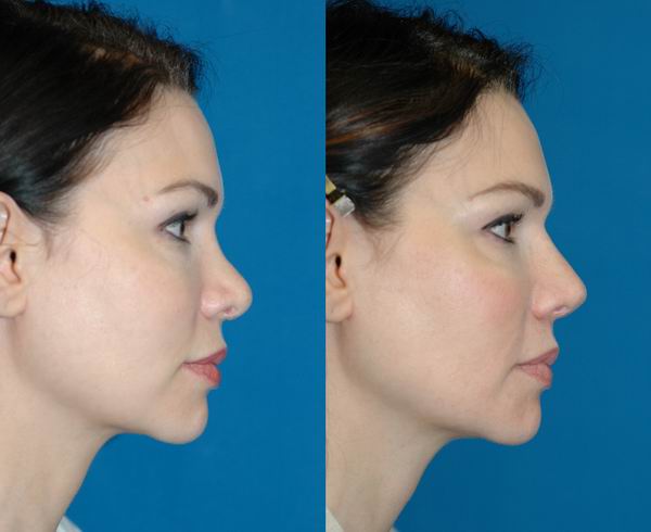
Revision rhinoplasty for polybeak deformity using rib cartilage graft by Dr. Sam Naficy. * Individual results may vary.
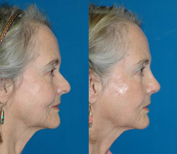
Revision rhinoplasty for polybeak deformity using rib cartilage graft by Dr. Sam Naficy. * Individual results may vary.
Revision Rhinoplasty for Crooked Nose
A crooked, asymmetric, or twisted nose is a common revision rhinoplasty problem. Crooked noses will sometimes appear more so in photographs or when smiling. A crooked nose will typically also have two different profiles (i.e. the right profile and the left profile are different). The nose may have crooked bridge, crooked tip, crooked nostrils, or a combination of all. Crooked noses often also have functional (breathing problems) in addition to the cosmetic problems. Many crooked noses also have a crooked or deviated septum and it is critical to correct the septal deviation at the same time as correcting the exterior of the nose so that the nose has a straight foundation (think of the septum as being a foundation on which the rest of the nose sits - if the septum is crooked many times so is the nose). Some crooked and twisted noses result from removal of excessive amounts of cartilage during the initial rhinoplasty so that the nose becomes unstable and tends to lean and twist during the healing process, especially if there is deviation of the septum which was not properly addressed. Nasal fracture can also result in a crooked nose. In children sometimes a fracture in childhood may be very subtle and not noticeable but as the child grows (and the nose grows) the degree of deviation increases. A crooked or twisted nose is treated at first by correcting any deviation of the septum. The other crooked or twisted structures of the tip and bridge are often treated by a combination of maneuvers such as trimming, suture contouring, and cartilage grafting to reshape and reconstruct the nose into a more symmetric shape. The cartilage that is used for grafting typically comes from the septum, the rib, the ear, or a combination. If the nasal bones are also crooked, osteotomies (controlled fracture of the nasal bones) will also need to be performed to correct the bridge.
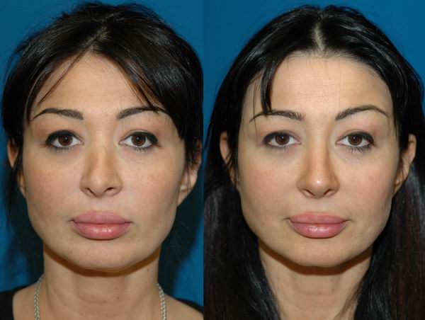
Revision rhinoplasty for crooked nose by Dr. Sam Naficy. * Individual results may vary.
Revision Rhinoplasty for Saddle Deformity
A saddle deformity or saddle-nose deformity results when there is inadequate support of the cartilage of the bridge of the nose, there area known as the middle vault. This loss of support results in collapse, settling or saddling of the mid portion of the bridge. A saddle deformity can be the result of over agressive reduction of the bridge and failure to add support to the middle vault during the original rhinoplasty. Saddle deformity can also occur from injury or fracture of the nose, especially if there is damage to the septum. Correction of a saddle deformity almost always requires cartilage grafting to rebuild the bridge of the nose. The cartilage grafts typically come from either the septum of the nose or from the rib.
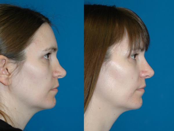
Revision rhinoplasty for saddle deformity of nose using rib cartilage by Dr. Sam Naficy. * Individual results may vary.
Revision Rhinoplasty for Hanging Columella
A hanging columella is when the center part of the nostrils under the tip is too droopy and is hanging down excessively resulting in too much nostril show. A hanging columella is at times also seen with alar retraction. Combination of alar retraction and a hanging columella makes the nostril opening seem excessively large. Correction of a hanging columella requires cartilage grafting from either the septum or rib to help support and straighten the columella.
Revision rhinoplasty for hanging columella using septal cartilage by Dr. Sam Naficy. * Individual results may vary.
Revision Rhinoplasty for Pinched Nostrils
Pinched nostrils are seen when there are 2 deep vertical lines (shadows) seen on either side of the tip, giving a pinched look to the tip and disrupting the continuity between the tip and the nostrils. Pinched nostrils are often caused by excessive removal of cartilage during the original rhinoplasty resulting in collapse of the weakened nostrils. Correction of a pinched nostrils requires cartilage grafting from either the septum,ear, or rib to help support the nostrils.
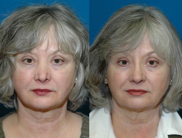
Revision rhinoplasty for pinched nostrils using rib cartilage by Dr. Sam Naficy. * Individual results may vary.
Revision Surgery for Nasal Valve Collapse
Nasal valve collapse (also known as nasal valve stenosis) is an anatomical abnormality involving the side walls the nose. When there is weakening and collapse of the nasal valves the side walls of the nose tend to cave in (collapse) when a deep breath is taken through the nose. Typically the reason for nasal valve collapse is that there is inadequate structural support (i.e. cartilage) in the side walls of the nose or that the cartilage is not shaped in the ideal way to keep the nasal passages open. In revision rhinoplasty, nasal valve collapse is seen in situations where too much cartilage was removed at the time of the initial rhinoplasty. Removal of too much cartilage weakens the side walls of the nose, causing them to collapse in under the weight of the skin. Noses with thick skin are more prone to nasal valve collapse since they need stronger cartilage support for their heavier skin. There are also a number of anatomical abnormalities of the nose cartilages that make the person at high risk for nasal valve collapse. The most common one is called cephalic malposition of the alar cartilages. In plain English this is a condition where the cartilages that are supposed to be supporting the side walls of the nose are not in the correct location and need to be surgically repositioned to provide support for the nasal valve. Repair of nasal valve collapse typically requires adding (grafting) pieces of cartilage to support areas of structural weakness and collapse in the side walls of the nose. The cartilage grafts usually come from the septum of the nose although they may also be taken from the rib or the ear.
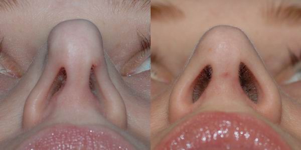
Revision surgery for nasal valve collapse using ear cartilage by Dr. Sam Naficy. * Individual results may vary.
Thick Skin in Rhinoplasty
Patients with thick (and sometimes oily) skin will often get underwhelming results after rhinoplasty if the surgeon uses the same techniques that are commonly applied for rhinoplasty in patients with thin or average skin thickness. Some may even feel that their nose is thicker/fatter after rhinoplasty. Thick skin conceals definition and contours of the underlying cartilages. To make matters worse, many patients with thick skin also have weak nasal cartilages. There are techniques for improving the shape of the thick-skinned nose and they involve a number of considerations:
- Keeping the structure of the nose strong
- Adding cartilage grafts to create more contour/definition to the nose
- When appropriate, thinning/trimming the fatty tissue from under the skin of the nose
Cartilage Graft Options
Most revision rhinoplasty surgery involves either rebuilding, support, or modifications of nasal cartilage. For this pupose cartilage is often added (i.e. grafted) to the nose. There are typically 3 choices of cartilage grafts used for revision rhinoplasty surgery.
- Septal cartilage obtained from the nose itself
- Ear cartilage obtained form the ack of the ear
- Rib cartilage obtained form the chest wall
Some surgeons may use sysnthetic implants or cadaver rib cartilage (cartilage obtained from a human organ donor) for revision rhinoplasty but we feel that these are not the optimal choice for grafting into the nose. Research studies have shown that implants and cadaver rib cartilage have a higher infection rate than when the patient's own living cartilage is used.
Septal Cartilage Graft
Septal cartilage grafting is almost always used in both primary and revision rhinoplasty. Septal cartilage is obtained from the septum (the midline wall of the nose) and it typically the first choice of grafting material.
Pros
- Septal cartilage grafts are typically available in noses that have not had prior surgery
- Septal cartilage grafts do not require any addtional incisions
- Septal cartilage grafts are natural and obtained from the patients own body
- Septal cartilage grafts coth firm to provide support and soft and pliable and make a good graft for the tip and sides of the nose
- Septal cartilage grafts are much more resistant to infection and absorption than implants or cadaver rib grafts.
Cons
- Septal cartilage is not always available in sufficient quantity needed for revision rhinoplasty
- Septal cartilage in some patients can be very thin, weak, and floppy and not suitable for situations where structural support is needed.
Ear Cartilage Graft
Ear cartilage grafting (also known as auricular cartilage grafting) is a traditional workhorse of revision rhinoplasty, especially when grafting is needed in the tip or alae (sides of nostrils). Ear cartilage is obtained from the back of the patient's ear at the same time as the revision. The procedure to harvest ear cartilage typically takes 10-15 minutes and requires placing an incision just over an inch on the back of the ear.
Pros
- Ear cartilage grafts are typically available where there may be inadequate donor cartilage in the nose
- Ear cartilage grafts are natural and obtained from the patients own body
- Ear cartilage grafts are soft and pliable and make a good graft for the tip and sides of the nose
- Ear cartilage grafts are much more resistant to infection and absorption than implants or cadaver rib grafts.
Cons
- Ear cartilage grafts require placing an incision behind the ear and the ear will be sore for a few weeks
- Ear cartilage grafts are not structurally strong and typically cannot be used to rebuild the entire bridge, lengthen a short nose, and correct severe deviations in cartilage
- Removing ear cartilage may at time create slight deformities of the ear.
Rib Cartilage Graft
Rib cartilage grafting (also known as costal cartilage grafting) is one of the workhorses of revision rhinoplasty. Patients presenting for revision rhinoplasty often have problems that require cartilage grafting but in many of those patients there is not adequate cartilage present in the nose to be used as grafting material. Rib cartilage obtained from the patient's own body at the same time as the revision rhinoplasty makes the ideal grafting material for a number of reasons. The procedure to harvest the rib cartilage typically takes 20-30 minutes and requires placing an incision just over an inch on the chest. In women the incision is hidden nicely in the inframammary fold.
Pros
- Rib cartilage grafts are typically available in large quantities when major grafting is needed
- Rib cartilage grafts are structurally strong and can be used to rebuild the entire bridge, lengthen a short nose, and correct deviations in cartilage
- Rib cartilage grafts are natural and obtained from the patients own body
- Rib cartilage grafts are much more resistant to infection and absorption than implants or cadaver rib grafts.
Cons
- Rib cartilage grafts require placing an incision on the chest and the chest muscle will be sore for a few weeks
- Rib cartilage grafts can feel somewhat hard and stiff for months although they do soften and feel more natural over time
- Rib cartilage grafts placed on the bridge of the nose can at times curve or 'warp' over time although there are technical modifications to avoid this
What Type of Anesthesia is Used?
A number of anesthesia options are available and your anesthesia provider will discuss with you which one is most appropriate for your health status and procedure. Some procedures require general anesthesia, while others may be done with IV sedation. With either, your heart rate, blood pressure, breathing and oxygen levels are monitored continuously by your anesthesia provider.
General anesthesia means that you are completely asleep for surgery and the placement of an intravenous line and a breathing tube is required. Frequently, numbing medication is also placed during surgery by your surgeon.
IV sedation is also called “monitored anesthesia care” or MAC. This involves receiving sedation and pain medication through an intravenous line (IV). At the beginning of the procedure, when you will be the sleepiest, your surgeon will be placing numbing medication in the area of the surgery. Once the area is numb you will require less sedation and pain medication but you will continue to receive enough medication to keep you sedated and comfortable during the entire procedure. During your surgery you may be receiving oxygen. Airway devices may be placed during IV sedation to keep you breathing normally.
Anesthesia guidelines [21kb PDF]
What is the recovery like?
A tape dressing will cover the nose for one week. With modern techniques employed by Dr. Naficy, no packing is required inside the nose. There may be some discoloration and swelling around the eyes which will improve over time. One week is usually enough time for returning to work and social activities although individual healing times may vary.
Post-operative care instructions [18kb PDF]
I am interested! What do I do next?
If you are considering this procedure we encourage you to complete this Surgical Consultation Intake Form. There is a great variety in nose shapes and features and each procedure must be custom tailored for the patient to get the best possible result. Dr. Naficy will tell you whether you are a suitable candidate for this procedure and inform you of the potential risks of the procedure.


